Table of Contents
ToggleEndosonography (EUS)
Although the most helpful endoscopic methods in the diagnosis of digestive system diseases, hidden masses and lesions may not be detected in some cases. However, by combining endoscopy and ultrasonography in a single device; diseases of the digestive system can be displayed. Thanks to this imaging, if necessary, a biopsy can be taken during the procedure and even sent for pathological examination.
Contents
What is EUS?
In which diseases is endosonography (EUS) used?
What are the advantages of endoscopic ultrasonography?
How is EUS done?
Frequently asked questions about endoscopic ultrasonography
What is EUS?
Endoscopy is a flexible instrument for examining the digestive system. Ultrasonography, on the other hand, is a device that allows images to be obtained from organs such as liver, gall bladder and pancreas with high-frequency sound waves. Endosonography, on the other hand, is a high-tech device that combines endoscopy and ultrasonography, allowing to examine the lower layers of the digestive system and surrounding tissues.
Endoscopic ultrasonography; Esophageal cancer, stomach cancer, rectum cancer, biliary tract cancer, pancreatic cancer diagnosis, biopsy, early detection of cancer, investigation of recurrence findings in postoperative follow-up, investigation of tumor size regression if radiotherapy was given before the surgery, in neighboring organs around the rectum ( prostate, bladder, vagina) is known to be significantly effective in examining cancer involvement in lymph nodes and vessels and determining the grade of the tumor (T1, T2, T3, T4).
In which diseases is endosonography (EUS) used?
Esophageal cancer, stomach cancer, rectum cancer, bile duct cancer, pancreatic cancer diagnosis, biopsy, early detection of cancer, investigation of recurrence findings in postoperative follow-up, cancer involvement in adjacent organs (prostate, bladder, vagina), lymph nodes and vessels around the rectum. examination and determination of the tumor grade (T1, T2, T3, T4)
Soft tissue tumors originating from the digestive system, submucosal lesions: Detection of tumors originating from the walls of organs such as the esophagus, stomach, rectum, gastrointestinal stromal tumor (GIST), leiomyoma, etc. detection of tumors
Biopsy of intra-abdominal lymph nodes
Anal fistula: Diagnosis and classification, understanding the relationship of fistula with breech muscles (anal sphincter)
Gas and fecal incontinence: assessing the thickness and condition of the anal canal muscles
Pancreatitis: Detection and evacuation of the pseudocyst fluid and necrotic tissues (in the case of necrotizing pancreatitis) around the pancreas called “pseudocyst” in cases of acute pancreatitis, evaluation of the pancreatic organ and duct in case of chronic pancreatitis
Pain management with EUS in pancreatic cancers
Biopsy from liver masses
Evacuation of bile ducts blocked due to cancer into the stomach or small intestine with EUS or stenting
What are the advantages of endoscopic ultrasonography?
The main advantage of endoscopic ultrasonography (EUS) is that it gives an idea about the deep spread of the cancer and its involvement in the surrounding lymph nodes, vessels and organs in cases such as esophageal cancer, stomach cancer, rectum cancer, pancreatic cancer and rectal cancer, and allows biopsy from these regions. .
Endoscopic ultrasonography (EUS) or endosonography is superior to standard ultrasonography in taking biopsy samples from lymph nodes and showing blood flow within the vessels.
Since endoscopic ultrasonography (EUS) or endosonography is performed with endoscopy, the interior of the organs is entered and thus images are taken much more closely and in detail.
With EUS, tissue samples can be taken from tumors originating from the digestive system and organs adjacent to the digestive system, cysts, etc. formations can be emptied.
Since EUS shows the wall layers in the digestive system in detail, it is used in the staging of tumors in the digestive system or in organs adjacent to the digestive system (the size and depth of the tumor, lymph node and adjacent organ metastasis, etc.) and in the examination of lesions located under the epithelium in the digestive system.
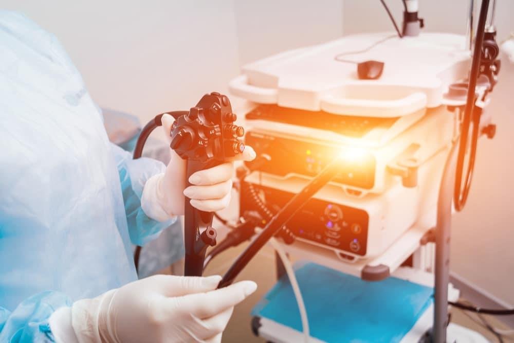
How is EUS done?
EUS procedure is not different from classical endoscopy. EUS can be performed both in the upper (oesophagus, stomach and duodenum) and lower digestive tract (large intestine). Preparation before the procedure, points to be considered after the procedure and possible side effects related to the procedure in EUS are not different from those in classical endoscopy. EUS may take longer than normal endoscopy. During the procedure, which takes an average of 30 minutes, the patient is given sedative and pain relievers. The procedure is carried out in an extremely comfortable way. Outpatients who come to the hospital for EUS procedure are usually sent home after being monitored for a few hours after the procedure and can return to their normal daily lives the next day.
Frequently asked questions about endoscopic ultrasonography
What should be considered before the procedure?
Before the procedure, the doctor must be informed about regularly used drugs such as aspirin, heart, high blood pressure and diabetes drugs, and current diseases. It may be necessary to stop the pressure from the drugs before the procedure.
What is the next process like?
It is recommended that the patient eat light and watery meals within the recommended period after the EUS procedure. The patient may experience burning and stinging for 1-2 days, so gargling with salt water is recommended. If problems such as severe vomiting, nausea and high fever are encountered after the procedure, it is recommended to consult a doctor.