Table of Contents
ToggleCardiac MRI
Cardiac MRI is a highly effective diagnostic method that provides detailed information on the type and characteristics of heart diseases without the need for interventional procedures, enabling rapid diagnosis of the disease and accurate treatment planning. It is among the preferred safe methods as it does not cause radiation exposure while providing high quality images.
Contents
What is cardiac MRI?
In which diseases is cardiac MRI used?
How is cardiac MRI done? / How is it applied? What kind of process awaits the person?
Frequently asked questions about cardiac MRI
What is cardiac MRI?
Cardiac MRI is an advanced cardiovascular imaging method that provides detailed images of the heart using radio waves, magnetic field and computer.
In which diseases is cardiac MRI used?
One of the most frequently used areas is heart failure. The degree of heart failure, changes in chambers and valves provide advanced information on whether heart failure is reversible or not. Apart from this, as mentioned above, pericardium (heart membrane), congenital heart diseases, valve diseases, aortic diseases; In coronary artery disease, the degree of involvement of the heart muscle and whether there is an oxygenation disorder are the areas that cardiac MRI can easily guide.
Cardiac MRI is also used in patients with palpitations, fainting, dizziness, and rhythm disturbances to investigate whether there is a focus or anatomical, hemodynamic cause. In patients with congenital heart diseases and aortic diseases, it provides a painless diagnosis without radiation, and helps in the follow-up and treatment process in patients with poor image quality and undiagnosed with echocardiography.
How is cardiac MRI done? / How is it applied? What kind of process awaits the person?
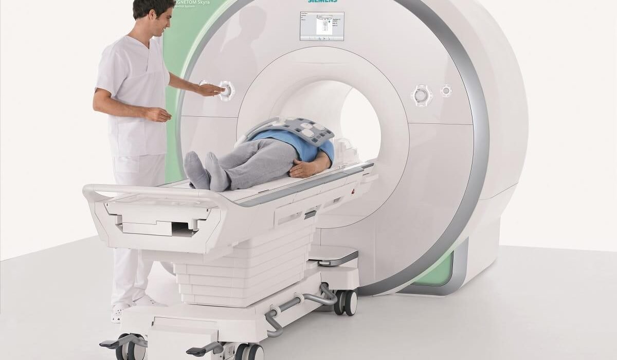
First, a detailed medical history was taken from the patients; Detailed information is obtained about the patient’s kidney functions, allergic conditions, pacemaker, whether there is any metal in the body and pregnancy status. Since the capture and image quality is very important for the correct interpretation of the examination, the shooting; It is a part of the process that needs to be studied very meticulously and requires extra care. First of all, vascular access is opened so that the contrast agent and other drugs that may be needed can be given to the patient. After the vascular access is opened, the patient is taken to a comfortable and wide cardiac MRI (Magnetic Resonance Imaging) machine. Cardiac MRI is a device with magnet feature, in the middle of which is a tunnel through which the patient bed enters. Electrodes that record the heart rate are attached to the patient’s chest. From the outside, the patient is told to breathe with voice commands. Each breath-hold time can usually last up to 10 or 20 seconds. In this process, it is very important for the image quality that the patient obeys the breathing commands and does not move at all. Make sure that you do this part correctly by making trials with the patient beforehand. The shooting takes about 50-60 minutes and there is no pain or suffering. Afterwards, the images are analyzed in detail and the reports are delivered to the patients quickly.
Frequently asked questions about cardiac MRI
Which patients should not undergo cardiac MRI?
Shooting is not applied to patients who have severe claustrophobia, i.e. fear of enclosed spaces, and do not agree to enter the device. In addition, if the urea and creatinine values in blood tests of patients with kidney failure are above a certain level, these patients should be evaluated by their physicians beforehand. General MR rules also apply to cardiac MR. It is not recommended if the patient has a metal prosthesis or a battery incompatible with MR anywhere in the body. Pacemakers or metal caps, if any, must be compatible with MRI scans. Before deciding on the procedure, the patient should be asked whether there is a metal prosthesis in the prosthetic cap, battery or any other part of his body. In addition, metals such as piercings need to be removed.
Is there a preparation process for cardiac MRI?
Although there is no special preparation process, it is important for the success of the examination that the patient shaves the hair on the chest, if any, before they come, due to the electrodes attached to the chest and recording the heart rhythm in the cardiac MRI procedure. In addition, if there is an allergic condition while using the pre-procedure drugs, 4-5 hours of fasting is required to minimize the risk of nausea-vomiting and aspiration of the patient. In addition, unless otherwise stated, the drugs used can continue to be used until the day of the examination. Bringing all the previous examination results and reports about the heart before the procedure will be beneficial for a more holistic examination of the patient.
What should be considered after cardiac MRI?
There is no condition that needs attention after the procedure. The patient resumes his daily life immediately after the procedure.
What are the advantages of cardiac MRI?
Unlike other cardiac imaging methods, echocardiography and cardiac tomography, the most important feature that distinguishes this method is that it provides detailed information at the tissue level. There is no other diagnostic method that can evaluate the tissue of the heart muscle in this way. For example, when echocardiography is performed on a patient with heart failure, only the presence and degree of heart failure is detected. However, when cardiac MRI is performed, very important information is obtained about the cause of heart failure. to heart failure; Cardiac MRI is the cardiac imaging method that can best answer the question of whether it is caused by coronary artery disease, a congenital genetic heart failure, or an inflammatory process due to infection in the heart. In this way, the diagnosis and treatment process of the patients is accelerated and a great contribution is made to the course of the disease.
While providing all these, it is a very important feature of this technique that it does not cause radiation exposure to the patients.
In addition to the diagnosis and treatment of the patients, it is also important to follow up the patients after the treatment process has started. For example, when a patient is evaluated with cardiac MRI before bypass surgery, the amount of tissue that can be healed after surgery can be predicted. Cardiac MRI can be used in follow-up to make detailed measurements and to evaluate on tissue basis.
In chemotherapy patients, some drugs can have a toxic effect on the heart tissue. Especially in patient groups whose echocardiographic image quality is not good, cardiac functions can be followed in detail with MRI. In addition, since there is no radiation, it is also of great importance for cancer patients.
Cardiac MRI can also be used to investigate whether there is any permanent damage after the treatment of heart muscle inflammation (myocarditis) or after a heart attack.
The heart muscle may also be affected due to some rheumatological diseases. If there is involvement in the heart, if heart failure is thought to be due to these diseases, the treatment can be completely changed. For this reason, cardiac MRI is very instructive in terms of whether there is involvement of the heart in rheumatological diseases.
The image quality of cardiac MRI is far superior to echocardiography. Detailed analysis can be made about the volume of the heart, the force of contraction and its entire anatomy. In particular, the right ventricle has a different, difficult, funnel-shaped anatomy, and while the detailed evaluation of the right ventricle is difficult with echocardiography, both volume and function can be easily evaluated with cardiac MRI. Cardiac MRI is one of the diagnostic criteria in the guidelines for heart disease called arrhythmogenic right ventricular dysplasia (ARVD), which can lead to life-threatening rhythm disturbances.
Since the connective tissue change in the heart muscle provides advanced information about possible rhythm disturbances, it is absolutely necessary to do this in some diseases with genetic origin, such as hypertrophic cardiomyopathy. In addition, since this disease has a genetic transmission pattern that we call ‘autosomal dominant’, when the family members are evaluated by echocardiography, if there are in-between conditions or early stage disease is suspected, it may be considered to control family members with cardiac MRI for detailed evaluation and evaluation on a tissue basis.
In intracardiac mass evaluation, it provides detailed information about the type of mass and its spread.
Since it provides advanced information in terms of anatomy, volume and hemodynamics in congenital (congenital) heart diseases, it is used quite frequently. It is a preferred diagnostic method in terms of being radiation-free, having the opportunity to evaluate hemodynamics and obtaining detailed images in the patients’ decision on the type of surgery and in their routine follow-ups.
How long does a cardiac MRI take?
It takes about 50-60 minutes.
Does cardiac MRI harm the kidneys?
If the patient’s kidney functions are impaired, hydration (intravenous serum administration) is performed or some measures are taken for this. If there is a very serious kidney problem, the process is carried out together with nephrology. Those with kidney problems should definitely notify the physician before cardiac MRI. The creatinine value is already checked routinely before cardiac MRI. If the patient has a recent creatinine value, it is not requested again. However, if it is not available soon, blood creatinine is checked again.
Does it have any side effects?
As mentioned above, cardiac MRI has no side effects, except for the possibility of allergy to the contrast material to be administered during the procedure and the progression of kidney disease in some kidney patients if care is not taken and precautions are not taken.
Is it routinely taken in the check up program?
If the patient does not have any indication, there is no need for a routine extraction. However, if the patient has a disease that requires follow-up such as heart failure, valve disease, congenital heart disease, aortic enlargement, it may be recommended to follow up and evaluate these patients with cardiac MRI. Especially in patients with aortic enlargement and who require close follow-up every year/2 years, follow-up with cardiac MRI instead of computed tomography will eliminate radiation exposure.
Is contrast material given in cardiac MRI?
Contrast material is given in cardiac MRI. There are conditions called late or early gadolinium uptake in cardiac MRI. In early involvement, it is checked whether there is any clot in the heart or a problem called microvascular obstruction. In the late gadolinium phase, contrast is required to see if there is a connective tissue change in the muscle tissue of the heart called myocardium. If the contrast agent is not given, it is not possible to see the connective tissue change in the heart muscle, the localization or the amount of dead heart cells.
