Table of Contents
ToggleWhat is Cranial Functional Magnetic Resonance Imaging (fMRI)
With its high resolution, Functional Magnetic Resonance Imaging (MRI) gives superior results compared to other imaging methods in imaging the anatomical structure of the brain and diseases. With this method, brain diseases can be evaluated in more detail. Cranial Functional Magnetic Resonance Imaging (fMRI), also known as Cranial Functional MRI, is an MRI method and provides significant advantages in the diagnosis and treatment of the disease. fMRI measures tiny changes in blood flow that occur with brain activity. fMRI enables the visualization of active areas by using the responses given by various stimuli and changes in blood flow and oxygen level in the brain tissue.
Contents
What is Cranial Functional Magnetic Resonance Imaging (fMRI)?
What does Cranial Functional Magnetic Resonance Imaging (fMRI) measure?
What are the uses of Cranial Functional Magnetic Resonance Imaging (fMRI)?
How is Cranial Functional Magnetic Resonance Imaging (fMRI) done?
What are the benefits and risks of Cranial Functional Magnetic Resonance Imaging (fMRI)?
Frequently asked questions about Cranial Functional Magnetic Resonance Imaging (fMRI)
What is Cranial Functional Magnetic Resonance Imaging (fMRI)?
Cranial Functional MRI is used to anatomically locate normal and diseased brain functions. MRI is a medical imaging method used in the diagnosis and follow-up of diseases in all body areas. Images are created using MRI, strong magnetic field and radiofrequency waves. MRI does not contain radiation. fMRI, on the other hand, is an MRI method that shows functionally activated areas in the brain depending on the change in blood oxygen level. fMRI provides visualization of functional areas of the brain that cannot be detected by other standard MRI imaging modalities.
What does Cranial Functional Magnetic Resonance Imaging (fMRI) measure?
Oxygen reaches neurons, which are brain cells, through the hemoglobin in the capillaries. When the activity of neurons in the brain tissue increases, the oxygen requirements also increase. In this case, blood flow increases in the area where neuronal activity increases. This situation also increases blood flow in the area where neuronal activity is increased.
Cranial Functional MRI provides images by measuring the changes in cerebral blood supply dependent on blood oxygen level of neurons in the brain at rest or during function. When neural activity increases in a certain area of the brain, the MR signal also increases slightly. fMRI identifies areas compatible with the activity by analyzing the MR signals obtained.
What are the uses of Cranial Functional Magnetic Resonance Imaging (fMRI)?
fMRI is used to anatomically locate normal and diseased brain functions. Although fMRI is used in stroke, trauma and some degenerative brain diseases, it is most commonly used in the clinic today to determine the relationship of the diseased area in the brain with critical functional structures before brain surgeries or other procedures to be applied to the cranial area (such as radiotherapy, gamma-knife therapy). The relationship between the diseased tissue in the brain (such as tumor, epileptic focus, vascular lesions) and the areas that provide speech, walking, hand-arm-leg movements and sensory functions are determined. fMRI is the preferred diagnostic method to evaluate the possible risks of brain surgery or other interventional treatments of the brain and to determine the most appropriate treatment method.
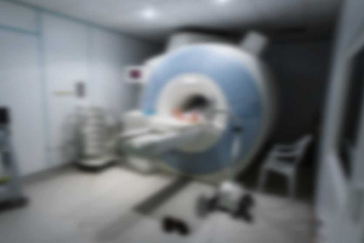
How is Cranial Functional Magnetic Resonance Imaging (fMRI) done?
For Cranial Functional MRI, the patient should lie still, as in other routine MRI examinations. An fMRI of the brain takes 30-40 minutes. If routine cranial MRI or cranial MR perfusion examination is not added during fMRI examination, MR contrast material is not used. In the MR room, there is a screen setup that the patient can easily see while lying on the MR device. With the help of the images projected on the screen, the patient is asked to perform various functions during the examination. The patients are informed in detail by the radiology specialist who will perform the examination before they enter the examination. As with all MRI examinations, it is important to keep the patient’s head still for best results. During Cranial Functional MRI, the patient is asked to perform operations that will enable us to identify functional areas in the brain while lying still. Images are obtained by asking the patient to perform simple operations such as hand, arm, leg, tongue movements, or to derive words from the mind with some methods. Exercises increase activity in certain areas of the brain. This activity allows the creation of a functional map of the patient’s brain with the images generated by the MR device. By doing these basic functions, it helps surgeons find the safest approach to removing a lesion, tumor from the brain, or surgery for epilepsy patients.
What are the benefits and risks of Cranial Functional Magnetic Resonance Imaging (fMRI)?
Cranial Functional MRI is a safe, painless and non-invasive imaging method. The most important advantage of fMRI is that it enables you to have a safe surgical procedure by detecting the areas of your speech and hand-arm-leg motor skills in the brain before the operation or interventional procedure. fMRIs help neurosurgeons prepare for brain surgery, allowing them to successfully reach the right area during surgery. As with routine MRI methods, fMRI is not a method that will harm you if you do not have a metallic or electronic device incompatible with the magnetic field. You will be asked to read and approve the information form and consent form before you are taken to the MR device.
Frequently asked questions about Cranial Functional Magnetic Resonance Imaging (fMRI)
How should I prepare for Cranial Functional Magnetic Resonance MRI?
Cranial Functional MRI examination and preparation before routine MRI examination are almost the same. The radiology technician starts preparing you for the examination by welcoming you. You may be asked to wear a hospital gown, or if your clothes are comfortable and metal-free, they may also be examined. It is recommended not to use the substance and make-up material applied to your face during MR imaging of your head and neck region, as they will distort the image. Jewelry and other accessories should not be on you. Watches, credit cards, hearing aids, pins, hairpins, metal zippers, removable teeth, pens, pocket knives and glasses, body piercings, cell phones, electronic items are not allowed in the MRI room. In most cases, patients with metal implants are safe for MRI, with the exception of a few types used specifically after 2000. However, people with some cochlear (ear) implants, brain aneurysm clip types, and some old pacemakers should not be taken into the MRI room without being evaluated for safety. If you have shrapnel, bullets or any of the other metals in your body, you must inform the radiology technician. In some special MRI examinations, there may be different applications related to eating and drinking and the drugs you use. Unless you are told otherwise, you do not need to be hungry and you can take your medication. Contrast material may be used in some MRI examinations. Contains MR contrast agent ‘gadolinium’. It can be used in patients with gadolinium iodine contrast allergy. A patient is much less allergic to gadolinium contrast than to iodine contrast. Before your MRI examination, you will be informed about whether you are allergic to contrast material, food or drugs. Be sure to indicate every question asked to you on the consent form and share it with the technician who prepared you. If you have any serious illness or surgery, tell the radiology technician who prepared you. Some conditions, such as advanced kidney disease, may require the use of certain types of gadolinium contrast that are considered safe for patients with kidney disease. In our department, we only use contrast agent derivatives that are stated to be safe. You may need a blood test to determine if your kidneys are working normally. If there is a possibility of being pregnant, it should be stated before the MRI examination. MRI has been used since the 1990s. No adverse effects on pregnant women or their unborn babies have been reported. However, MRI is not recommended for pregnant women in the first trimester unless the benefit of the procedure outweighs any potential risk. MR contrast material should not be used in pregnant women unless necessary.
Should I come hungry or full for Cranial Functional Magnetic Resonance MRI examination?
The radiology technician will make a detailed interview with you before the extraction. Unless you are told otherwise, you do not need to be hungry, you can do your usual routine nutrition.
Can I drink the drugs I use for other diseases before the Cranial Functional Magnetic Resonance MRI examination?
In some special MRI examinations, there may be different applications related to eating and drinking and the drugs you use. However, in other cases, you can take your medicines unless you are told otherwise.
Is there Cranial Functional Magnetic Resonance MRI radiation?
Cranial Functional MRI does not contain radiation.
Can I have Cranial Functional Magnetic Resonance MRI when I am 3 months pregnant?
First of all, female patients who are likely to be pregnant should confirm this possibility by performing the necessary pregnancy tests before the Cranial Functional MRI examination. No adverse effects on pregnant women or their unborn babies have been reported. However, it is not recommended for pregnant women to have fMRI in the first trimester (3 months of pregnancy), unless the benefit of the procedure outweighs any potential risk.
Is contrast material used in pregnant patients in Cranial Functional Magnetic Resonance MRI?
Cranial Functional MRI can be performed if necessary for patients who have completed the 3-month pregnancy period. In addition, MR contrast material should not be used in pregnant women unless it is mandatory.
There is a genetic kidney disease in my family, can I have Cranial Functional Magnetic Resonance MRI?
Patients who have a serious illness or have undergone any surgery must report this to the radiology technician. Some conditions, such as advanced kidney disease, may require the use of certain types of gadolinium contrast that are considered safe for patients with kidney disease. MR contrast agents used in our hospital are in the safe group. You may also be asked for blood tests to determine if your kidneys are working normally.
Should I remove my body piercings before Cranial Functional Magnetic Resonance MRI?
Jewelry and other accessories should not be on you. Watches, credit cards, hearing aids, pins, hairpins, metal zippers, removable teeth, pens, pocket knives, glasses, body piercings, mobile phones and all electronic items are not allowed in the MR room.
Can I have a Cranial Functional Magnetic Resonance MRI examination with my own dress?
It is preferred that you enter the MRI room with a hospital gown for fMRI examination. However, if your clothes are comfortable and do not contain metal, they can also be examined.
Do I need to remove my makeup before Cranial Functional Magnetic Resonance MRI?
It is recommended not to use the substance and make-up material applied to your face during MR imaging of your head and neck region, as they will distort the image.